POST PROVIDED BY CHRIS BROECKHOVEN, ANTON DU PLESSIS, STEPHAN G. LE ROUX, P. LE FRAS N. MOUTON AND CANG HUI
X-ray micro-computed tomography – or µCT – is a technique that uses x-rays to create high resolution cross-sections of samples. Virtual 3D models can be made from these cross-sections without destroying the original samples. Micro-CT has important applications in medical imaging and, in the biomedical field, in vivo µCT allows researchers to make virtual 3D models of the skeleton and organs of live small animals. Three-dimensional models like these could provide insight into diseases and guide the development of medicines and therapies.
In vivo µCT holds three major advantages over other methods:
- It allows for repeated measurements of small live animals at different times without having to sacrifice them.
- It eliminates variation among individuals.
- It can reduce the number of animals required to obtain statistically meaningful data.
A variety of commercially available µCT scanners that are optimised for scanning live animals are now available. The use of in vivo µCT in ecological and evolutionary studies, however, has greatly lagged behind its use in biomedical studies.
Reptiles and amphibians, especially lizards, have a long history of serving as model study organisms for an array of ecological and evolutionary studies, but invasive techniques still dominate morphological measurements of skeletal features. Micro-CT scanning has the potential to significantly advance this field, but there are issues that need to be addressed first.
Overcoming the Hurdles of In Vivo µCT in Non-Medical Studies

The main barrier hindering the advancement and implementation of in vivo µCT is the limited research access to commercial µCT scanners optimised for small living animals. Various scientific institutions however, such as the CT scanner facility at Stellenbosch University (South Africa), are in the possession of a different type of µCT scanners, often referred to as industrial µCT scanners. Industrial µCT scanners differ from the commercial variety in that the sample holder rotates while the X-ray source and detector remain relatively stationary, rather than the X-ray source and detector rotating around a stationary sample (similar to a clinical CT scanner used in hospitals).
There are two major issues with this rotating sample design:
- There is very limited information is available on the radiation doses produced by industrial µCT scanners that are not developed for in vivo animal studies.
- The design would be incompatible with equipment required during anaesthesia. In addition to this, the administration of anaesthesia in amphibians and reptiles itself is often problematic and requires practical and ethical considerations.
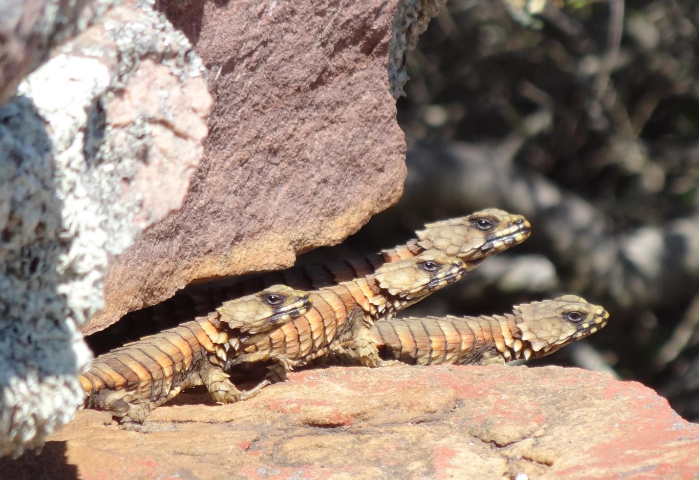
The development of an in vivo µCT protocol for reptilian and amphibian models, strongly depended on whether we could overcome these two major issues. In ‘Beauty is more than skin deep: a non-invasive protocol for in vivo anatomical study using micro-CT’, recently published in Methods in Ecology and Evolution, we present an inexpensive, non-invasive solution to both problems. We use an examination of bones in the South African armadillo lizard (Ouroborus cataphractus) as an example.
A Brief Overview of the In Vivo µCT Protocol
Our protocol deals with the two disadvantages of industrial µCT scanners.
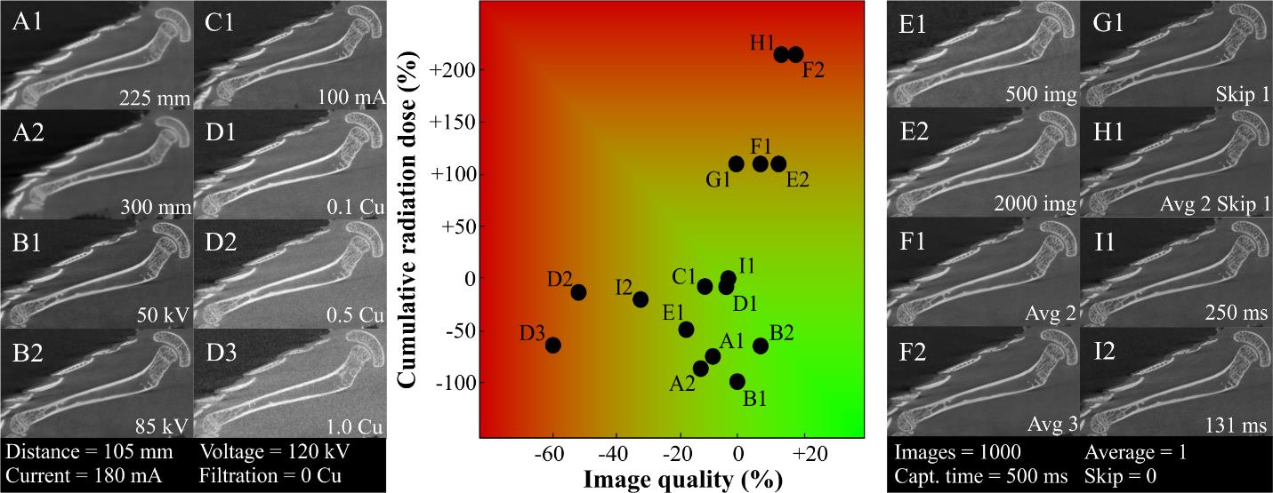
Radiation dose – Exposure of animals to radiation is a major concern for µCT imaging. We conducted a number of experiments, using a museum specimen, to investigate the effect of various scanner parameter settings on radiation dose and image quality. By adjusting specific parameters, such as x-ray tube voltage or distance of the sample to the x-ray source, we found that radiation dose could be greatly reduced without compromising image quality. In fact, the radiation doses produced in our protocol were similar to, or even lower than, those reported by studies using µCT scanners optimized for in vivo imaging.
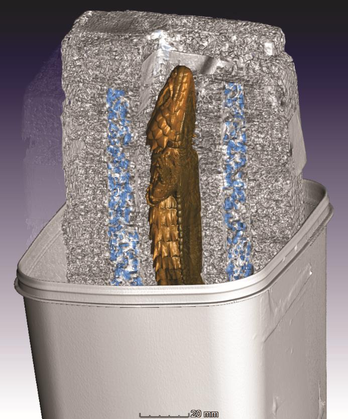
Immobilisation and restraint – Reptiles and amphibians hold one great advantage over mammals: they are cold-blooded. This means that cooling can be used as a method of immobilisation. Cooling is more cost-effective and practical than the administration of anaesthetics and does not cause additional stress or risks to the study organism. Lizards were cooled to ± 8°C using a portable incubator placed in the CT-scanning facility to allow for direct transfer from incubator to the µCT scanner. To restrict movement, lizards were restrained between two Styrofoam plates and placed in a custom-built Styrofoam holder. A layer of crushed ice was placed between each side of the holder and the plates restraining the lizard.
Lizards remained near immobile for the entire duration of the scan, which takes up to 16 minutes, without the need for anesthesia. By using cooling as immobilization method, we acquired a comparative image quality of in vivo scans compared to that of scans using a museum specimen. Our only caveat was that breathing resulted in a slight blur in the lung region, an issue that further improvements will definitely overcome.

Potential Applications of In Vivo µCT for Ecological and Evolutionary Studies
The proposed in vivo µCT scanning protocol offers ecologists and evolutionary biologists several benefits including repetitive measurements of anatomical features, as well as accurate measurements of smaller structures which can be easily examined in detail at high resolution without having to sacrifice study organisms.

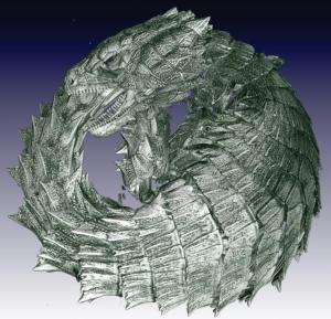
In addition, our µCT protocol holds another great advantage: specific behaviours could be investigated in vivo in the absence of anaesthesia. For example, armadillo lizards have a defensive tail-biting strategy which they use when threatened by a predator. This strategy involves rolling up into a ball and biting their own tails. In vivo µCT allowed us to obtain a high-resolution virtual 3D model of this unique behaviour which could provide new insights into how defensive morphologies can serve to protect against predators.
Industrial µCT systems are becoming widely available to ecologists and evolutionary biologists. While the biomedical studies that employ in vivo µCT technology are advancing rapidly, future studies should focus on improving techniques and protocols that make use of these more commonly available systems.
To find out more about in vivo µCT scanning, read our Methods in Ecology and Evolutionarticle ‘Beauty is more than skin deep: a non-invasive protocol for in vivo anatomical study using micro-CT’.
