
Post provided by Karsten Vesterholm
I’m a Postdoc in the Sound and Behaviour research group at the Department of Biology at the University of Southern Denmark, where I work in the Bat Echolocation Lab. We are particularly interested in understanding how bats use directional hearing as part of their echolocation. Direction of hearing is determined primarily by the shape and orientation of the outer ear (pinna) and the shape of the head and torso depending on the frequency of the sound.
These shapes filter incoming sounds differently depending on which direction sounds come from and is defined as the head-related-transfer-function (HRTF). The HRTF is very important when trying to understand how animals localize sound, doubly so for animals like bats that primarily navigate and forage using sound.
Traditionally HRTFs are either measured using an in-ear microphone with a vast array of surrounding speakers or numerically modelled based on 3D models obtained from µCT scans. Both these methods are very time consuming, require motion less (often dead) animals, and a µCT scanner is very expensive.
Seeing how ears hear
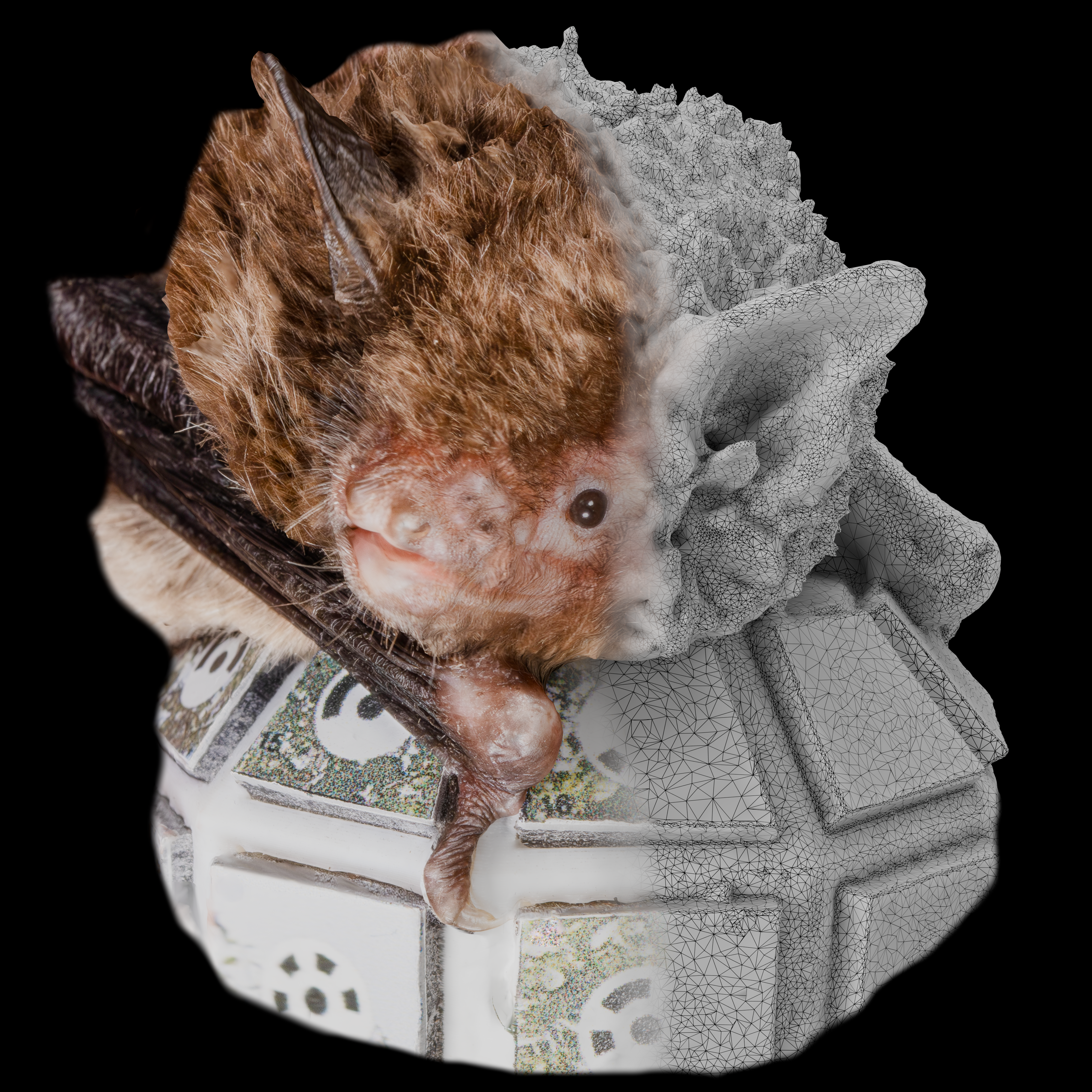
Photogrammetry is the process of capturing a set of overlapping images of the same subject and stitching the images together to form a 3D model of the subject similar to the µCT scan, but it can be done with a single camera.
Most importantly, with enough cameras, all data can be collected in the time it takes to capture an image which can be very fast, down to a few milliseconds or less. The 3D model of the animal is then used to simulate the HRTF using the standard numerical method called Boundary Element Method (BEM).
Photogrammetry therefore allows us to capture 3D models from alive and awake animals, and it lets us literally see how ears hear since our data comes from images.
If it works for bats, it works for anything
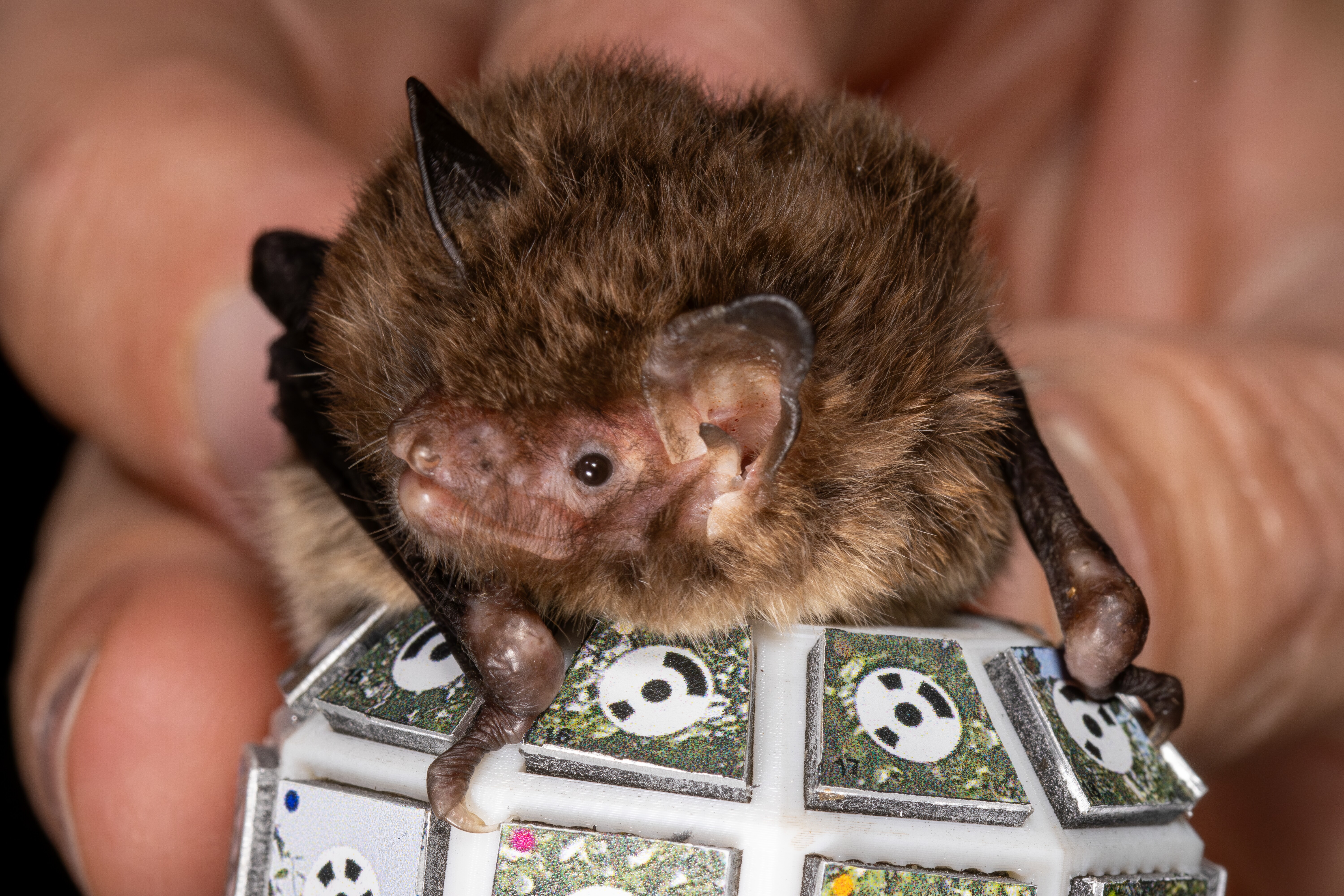
We consider bats to be the most challenging subject for this method. Bats are very small (ears can be as small as 1-2 cm), which in itself is a huge challenge for 3D photogrammetry. The biologically relevant frequencies go up to 100 kHz and beyond for some bat species, which is very demanding in regard to how accurately we can capture the shape of the pinna. After we had the method working for bats, we confirmed the general applicability by trying it out on a pig, where it also works.
Studying hearing as it actually happens
This method allows us to study the directional hearing of animals, while they are alive and awake. Ultimately, we envision this method will allow us to capture the ear shape of bats inflight in a lab setting.
No sacrifice and no anaesthesia
Since we no longer need the animal to be completely still for the scan, there is no need to sacrifice the animal or even use anaesthesia. This makes the method far very attractive since it allows us to quickly collect data from a vast number of individuals and to investigate rare and endangered species that would have been unavailable using previous methods.
Read the full article here.
