
Throughout March, we are featuring articles shortlisted for the 2025 Robert May Prize. The Robert May Prize is awarded by the British Ecological Society each year for the best paper in Methods in Ecology and Evolution written by an early career author. Or Ben-Zvi’s article ‘The Benthic Underwater Microscope imaging PAM (BUMP): A non-invasive tool for in situ assessment of microstructure and photosynthetic efficiency‘ is one of those shortlisted for the award.
About the paper
What is your shortlisted paper about, and what are you seeking to answer with your research?
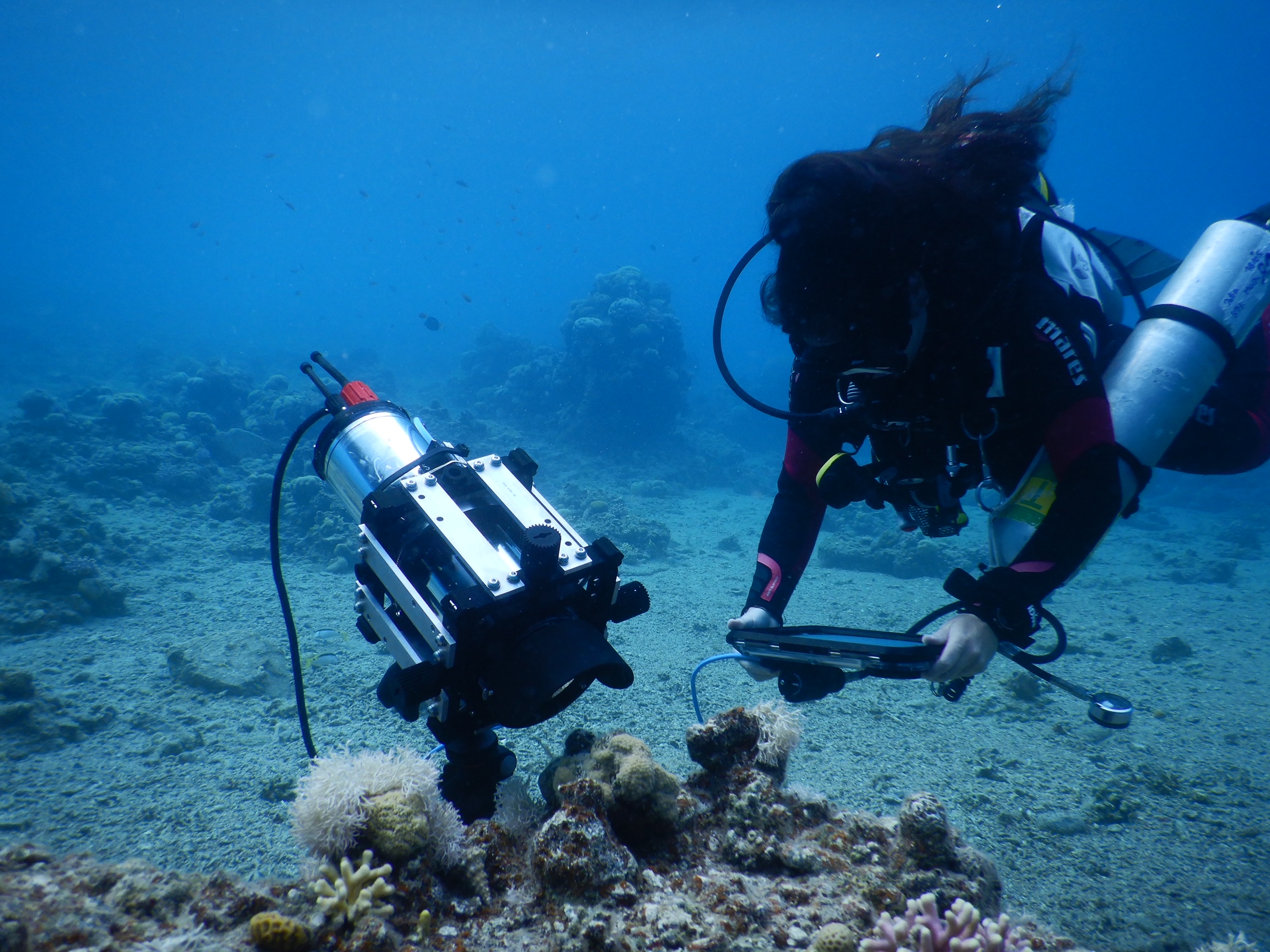
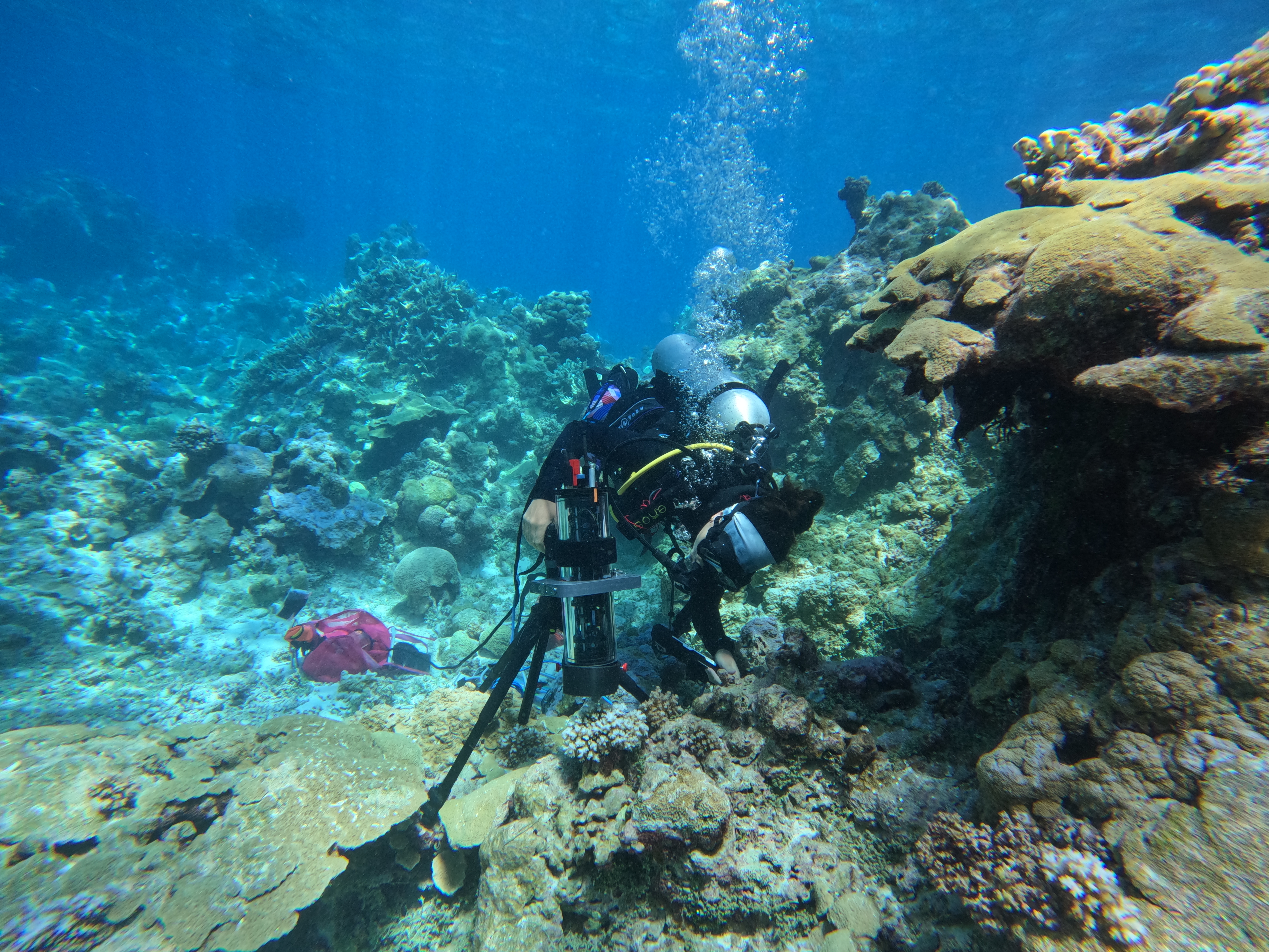
In this paper, we introduce a new underwater microscope with integrated photosynthetic efficiency measurements, the BUMP, and some of its potential applications. This instrument was created in collaborative work between engineers, physicists, and marine biologists to bring advanced imaging technology into coral reef research. We designed it to help scientists study micro-scale processes that drive large-scale changes in aquatic ecosystems. Specifically, we use it to investigate the photosynthetic capacities of symbiotic microalgae residing in coral tissue by observing corals at the scale of individual polyps while simultaneously measuring the photosynthetic performance of their symbiotic microalgae.
Were you surprised by anything when working on it? Did you have any challenges to overcome?
Working underwater at microscopic resolution is inherently challenging. Water motion and limited air supply make it difficult to stabilize and focus on tiny, living subjects. I was therefore pleasantly surprised by how stable the instrument proved to be in the field. One of the most exciting moments came when analyzing the images: coral polyps are far more dynamic than we often assume. Seeing them constantly moving and interacting with their environment was both scientifically illuminating and personally thrilling.
What is the next step in this field going to be?
For the microscope itself, I believe it opens the door to addressing key questions about coral–algae symbiosis and the mechanisms underlying bleaching. More broadly, I expect the field to move toward long-term, autonomous observations rather than diver-operated systems. Continuous monitoring at high spatial resolution will allow us to capture transient stress events and better understand how physiological thresholds are crossed in real time.

What are the broader impacts or implications of your research for policy or practice?
The microscope is a tool; its impact depends on how we use it. By uncovering the mechanisms that drive bleaching at the microscale, we can provide stronger mechanistic foundations for conservation and management strategies. In addition, the visual power of the images we produce is significant. High-resolution fluorescence images can help communicate both the beauty of corals and the fragility of their symbiosis, strengthening public engagement and support for reef conservation.
About the author
How did you get involved in ecology?
I was mentored by Prof. Yossi Loya, whose enthusiasm for coral reefs was contagious. His passion for asking fundamental ecological questions in the field shaped the way I approach science and inspired me to pursue marine ecology and later on, physiology.
What is your current position?
I am currently a postdoctoral researcher at Scripps Institution of Oceanography at UC San Diego.
Have you continued the research your paper is about?
Yes. I am continuing to investigate coral bleaching, focusing on disentangling the roles of each partner in the breakdown of the symbiosis. By combining physiological measurements with high-resolution imaging, I aim to better understand how stress responses unfold at the cellular and polyp scales.
What one piece of advice would you give to someone in your field?
Stay attentive. Some of the best ideas begin as casual observations. If you truly watch your study organism and its surroundings, unexpected patterns can emerge—and those moments often lead to the most meaningful discoveries.
