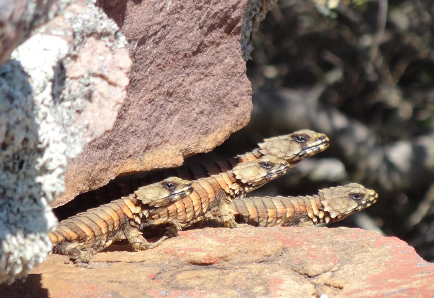
Post provided by Dylan Wainwright Our recent Methods in Ecology and Evolution paper – ‘Imaging biological surface topography in situ and in vivo‘ – shows how to use gel-based profilometry to image various biological surfaces. To start you need to press a gel into a surface of interest. The bottom surface of the gel is coated in a paint to create an impression of the surface that has standard optical … Continue reading Scales, Slime and Dragon(fly) Wings? Investigating the Surfaces of Organisms
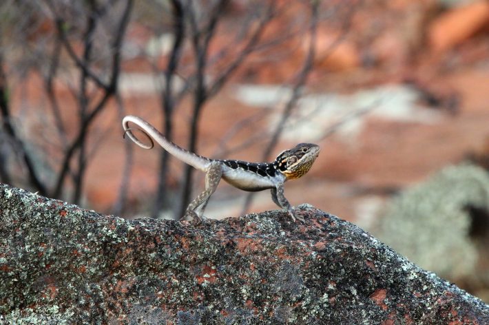
Many animals rely on movement to find prey and avoid predators. Movement is also an essential component of the territorial displays of lizards, comprising tail, limb, head and whole-body movements.
For the first time, digital animation has been used as a research tool to examine how the effectiveness of a lizard’s territorial display varies across ecological environments and conditions. The new research was published today in the journal Methods in Ecology and Evolution.
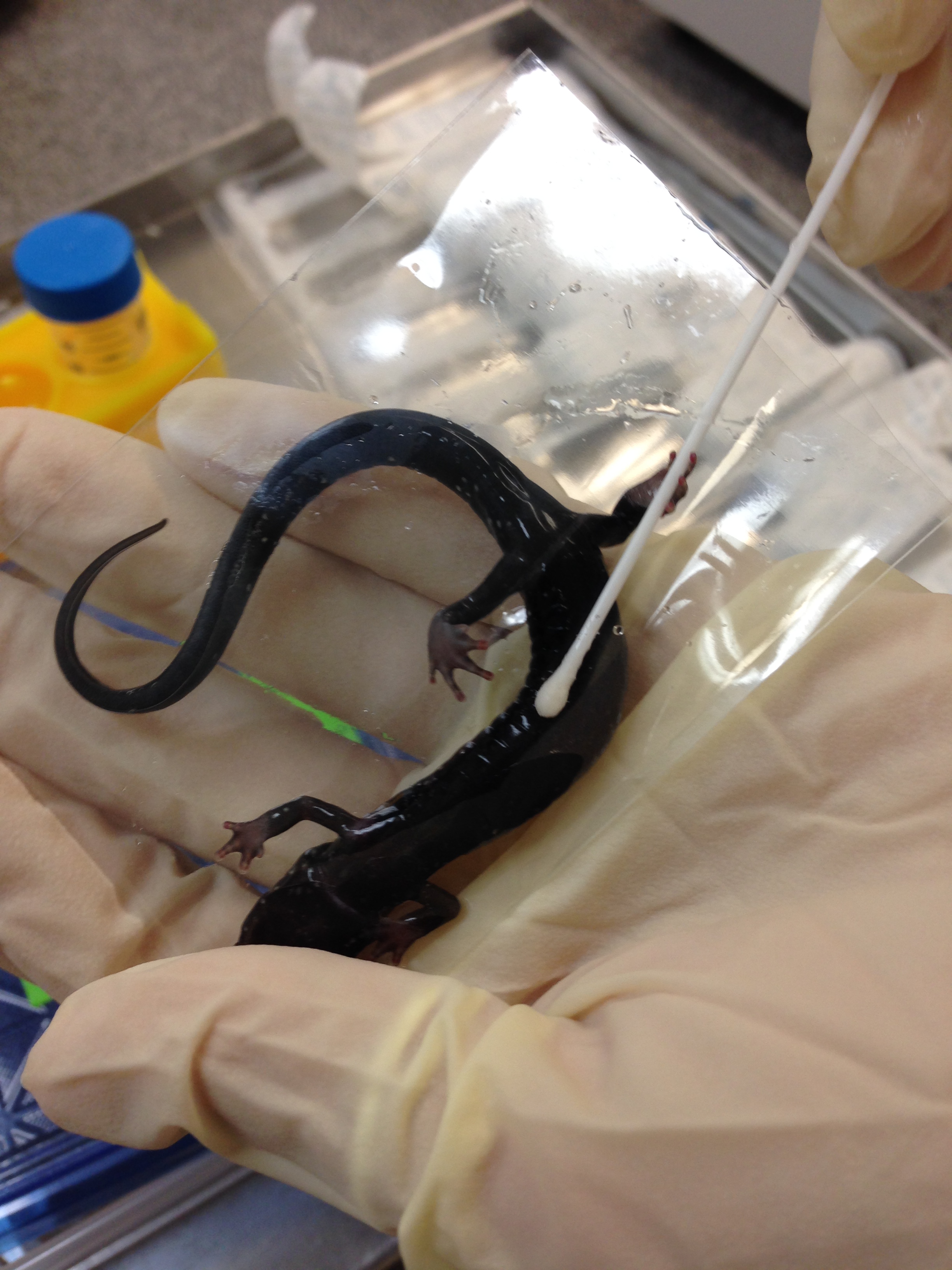
A salamander having its skin swabbed to test for Bsal infection.
Imagine you’re at the doctor’s office. You’re waiting to hear back on a critical test result. With recent emerging infectious diseases in human populations, you are worried you may be infected after a sampling trip to a remote field site. The doctor walks in. You sit nervously, sensing a slight tremble in your left leg. The doctor confidently declares, “Well, your tests results came back negative.” In that moment, you let out a sigh of relief, the kind you feel throughout your body. Then, thoughts start flooding your mind. You wonder– what are the rates of false negatives associated with the test? How sensitive is the diagnostic test to low levels of infection? The doctor didn’t sample all of your blood, so how can they be sure I’m not infected? Is the doctor’s conclusion right?
Now, let’s say I’m the doctor and my patient is an amphibian. I don’t have an office where the amphibian can come in and listen to me explain the diagnosis or the progression of disease − BUT I do regularly test amphibians in the wild for a fatal fungal pathogen, known as Batrachochytrium dendrobatidis (commonly known as Bd). Diseases like Bd are among the leading causes of the approximately one-third of amphibian species that are threatened, near threatened, or vulnerable to extinction. To test for Bd, and the recently emerged sister taxonBatrachochytrium salamandrivorans (hereafter referred to as: Bsal), disease ecologists rely on non-invasive skin swabs. Continue reading “Imperfect Pathogen Detection: What to Do When Sampling and Diagnostic Tests Produce Inaccurate Results”
Many researchers, breeders and hobbyists need to know sex of their animals. Sometimes it’s easy – in sexually dimorphic species you only have to look. In other species or juveniles it’s often not so straightforward though. And it’s often impossible – but sometimes essential – in embryos or in tissue samples. Determining sex from DNA is the most practical option, or sometimes even the only possibility, in these cases.
Molecular sexing is routinely used in mammals and birds, but until now it has only been available for a handful of reptile species. Many people didn’t believe that this situation would improve considerably any time soon. But why? Continue reading “Reptile DNA Sexing: Easier Than You Ever Thought”
Post provided by Vasco Elbrecht Many things can negatively affect stream ecosystems – water abstraction, eutrophication and fine sediment influx are just a few. However, only intact freshwater ecosystems can sustainably deliver the ecosystem services – such as particle filtration, food biomass production and the supply of drinking water – that we rely on. Because of this, stream management and restoration has often been in the … Continue reading Building Universal PCR Primers for Aquatic Ecosystem Assessments
X-ray micro-computed tomography – or µCT – is a technique that uses x-rays to create high resolution cross-sections of samples. Virtual 3D models can be made from these cross-sections without destroying the original samples. Micro-CT has important applications in medical imaging and, in the biomedical field, in vivo µCT allows researchers to make virtual 3D models of the skeleton and organs of live small animals. Three-dimensional models like these could provide insight into diseases and guide the development of medicines and therapies.
In vivo µCT holds three major advantages over other methods:
It allows for repeated measurements of small live animals at different times without having to sacrifice them.
It eliminates variation among individuals.
It can reduce the number of animals required to obtain statistically meaningful data.