Post provided by Jordan Cuff and Maximillian Tercel
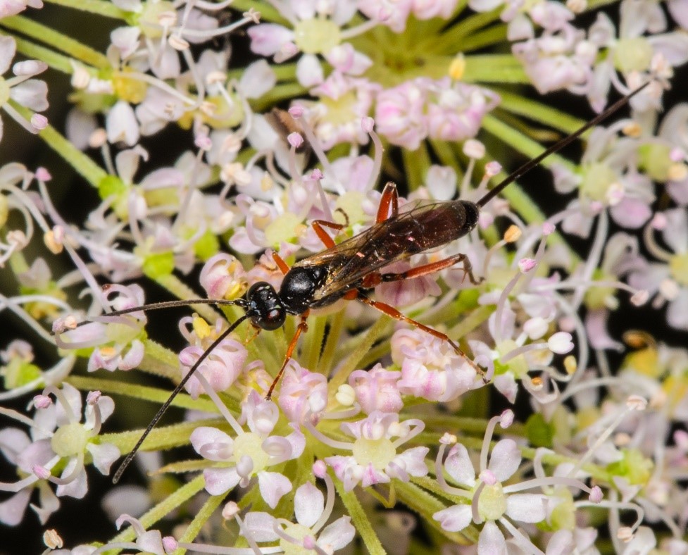
Are you kept awake at night wondering how you would measure the macronutrient content of small invertebrates? Perhaps you have tried but are haunted by the disappointment that you have had to rely on conversion factors, analogues and pooled samples. Get ready to sleep soundly, entomological entrepreneur!
In this blog post, Jordan Cuff and Maximillian Tercel will discuss their latest study published in Methods in Ecology and Evolution, concerning their brand-new method for measuring macronutrient content in invertebrates: MEDI.
Current challenges
Nutrient analysis of small specimens like invertebrates has always been constrained by a multitude of limitations, not least the insufficient sensitivity of most techniques to such small volumes of material. Therefore, many ecologists tend to use techniques which indirectly measure macronutrients through measuring analogues, such as nitrogen instead of protein. This approach then requires calculation of correction factors which often vary between taxa, making multi-taxon studies laborious at best. Direct measures do exist, and even for samples as small as tiny flies or adorable aphids, but they need one specimen per macronutrient, making individual comparisons impossible.
MEDI: Macronutrient Extraction and Determination from Invertebrates

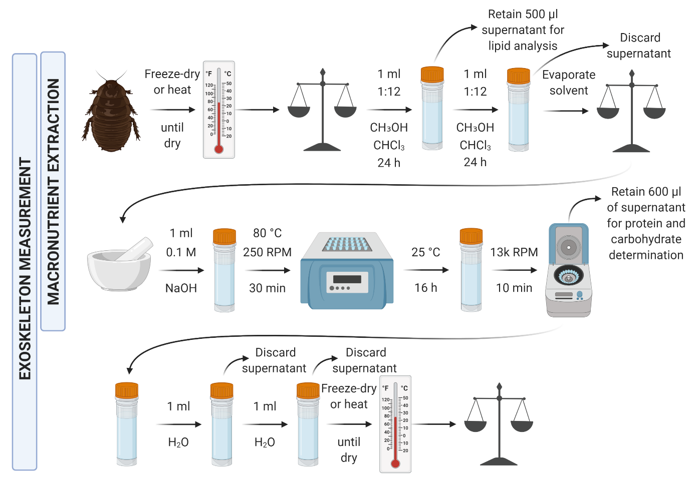
MEDI marks an important step forward for ecological nutrient analysis since it allows the direct measurement of three macronutrients from a single sample as small as a springtail (or perhaps even smaller). This is achieved by bringing together a two-step extraction procedure and three existing colorimetric assays. MEDI uses three (or more) different colorimetric assays to determine the content of protein, lipid and carbohydrate from a single specimen. Colorimetric assays basically allow us to measure how much of a target molecule is present by using chemical reactions to change the colour of the solution it is in, which we can then quantify by measuring the absorbance of particular wavelengths of light.

In our study, the Pierce modified Lowry protein assay, involves the measurement of blue light absorbance as the protein present reacts with the reagents to form a blue molecule (heteropolymolybdenum Blue). Importantly, colorimetric assays will often be biased toward or exclusive to a subset of the target macronutrient, but they will still provide an accurate means for direct detection and concerned users could mitigate these effects by combining multiple methods. These assays are rapid, relatively cheap and involve fairly standard laboratory equipment. The different macronutrients do, however, require different extraction methods.
MEDI uses a multi-step extraction protocol to allow the separate detection of lipids, and protein and carbohydrate. To extract lipids, MEDI uses a chloroform/methanol solution, which penetrates the tissue and solubilises the lipids present, the resultant solution being suitable for lipid analysis. Once this is removed (and any remnants evaporated) the specimen can then be lysed in sodium hydroxide (NaOH), which forms a solution containing the protein and carbohydrate constituents of the specimen. These two solutions can then be analysed using the above colorimetric assays to determine the concentration of macronutrients in these solutions (and thus the specimens). The concentrations are calculated with reference to a serial dilution of a standard for each macronutrient.
MEDI applications beyond invertebrates
The main focus of MEDI during its development was on invertebrates, but the protocol brings together methods relevant to a wide range of materials, so why stop there? Already, researchers have shown interest in applying the protocol to leaves, seeds, algae and tissue samples. The detectable sample size could also be made even smaller by changing the volume of reagents, incubation times or standard dilution series. The flexibility of the protocol could also allow inclusion of other assays for detecting micronutrients or other chemical groups.
Our study briefly compares several different analytical techniques for macronutrient analysis, and practically compares two protein assays. The purpose of this was to illustrate to readers and potential future users, that there is not one single catch-all method for the MEDI approach. For example, the Bicinchoninic acid assay (BCA) and Lowry assays were both used and briefly compared for protein measurement. In the case of the German cockroach Blattella germanica samples tested, the BCA assay was inaccurate since uric acid, which accumulates in these cockroaches, interferes with the assay, whereas this assay performed well with all of the other species tested. Ultimately, one could simply use the extraction and assay methods included in the study “straight out of the box” and likely see great results, but it is never a bad idea to first think about the focal material and how different assays or standards may suit it.
Ultimately, MEDI shows great promise as a widely applicable protocol for invertebrate (and beyond) nutrient analysis. The cost per sample calculated during this project was £1.21 (or $1.35), making this a relatively cheap and accessible method, taking into consideration slight fluctuations in this price due to material costs. We hope that it provides a great resource to the ecological research community and we look forward to seeing the incredible research that it is applied to going forward.
To read the full study published in Methods in Ecology & Evolution, ‘MEDI: Macronutrient Extraction and Determination from invertebrates, a rapid, cheap and streamlined protocol’ accepted in Methods in Ecology & Evolution, visit the journal website here.
