
Post provided by Aaron T Irving, Justin HJ Ng and Lin-fa Wang
Bats. They’re amazing creatures. Long-lived (with relevance to their body size), echolocating (for microbats and some megabats), metabolically-resilient (apparently resilient to most virus infections) flying mammals (with heart beats up to 1200 bpm for hours during flight). There are 1,411 species of this incredible creature. But very little is known about their physiology and unique biological traits. And detailed evolutionary analysis has only just begun.
The problem is, they’re an ‘exotic’ animal (wildlife that most people do not come into contact with). Being a long-lived animal producing minimal offspring (most only have one baby per year), they’re not suited to the kind of experimental studies we do with other animals like mice. Unavoidably, some aspects of biology require the use of tissues and cells. These samples can be used for sequencing, genomics, molecular evolution studies, detailed transcriptomic analysis, functional experiments with specific cell types and much more. Some methodology is beginning to be published – such as capture techniques and wing punch/genomic isolation – but there’s been an absence of protocols for the processing of bats. This is essential for the field to maximise the potential application of each individual and for minimising non-essential specimen collection.
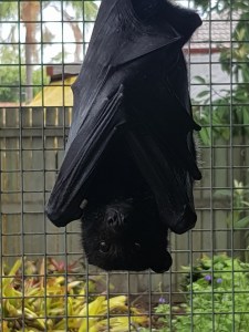
Access to the Australian Black Flying Fox
Invasive sample collection is important for molecular work, but we were cautious about impacting the natural populations of flying foxes. These large fruit bats reside in roosts, sometimes close to areas of urban expansion, and are susceptible to ‘re-settlement’ if they’re seen to be pests. While the IUCN states this species as ‘least concern’, there’s limited information on their population size and dynamics. This is largely because of their transient nature, wide-ranging distribution and limited support for population monitoring. The Australian black flying fox does have reliable genetic data available though. This includes an annotated genome, established cell lines for validation and transcriptomic data available for analysis. It’s also a large bat, making the process of sample collection much easier (and harder at the same time, their claws are huge!).
With our collaborators and their network of contacts we were fortunate enough to gain access to individuals already removed from the wild population, minimising the impact. These poor bats had been physically damaged beyond repair (mostly from barbed wire fences or powerlines) or were orphaned, abandoned, or dehydrated bats that couldn’t recover enough for release. They were scheduled for euthanasia due to laws imposed on the number of bats animal carers may house. The carers do an amazing job at rescuing most bats, but sometimes they’re overwhelmed by the sheer numbers.
We used this opportunity to collect samples and optimise sample collection from the animals that could not be saved. Our plan was to build a resource of cells and tissues for a variety of future experiments. We wanted to bank as many cell types as possible and have tissue for RNA, DNA and Protein, along with sera, urine, faeces, stomach contents etc. With the help of skilled veterinarians, we perfected dissection through surgical necropsy and identified the workflow and logistics to facilitate both speed and sample quality.
Finding a Facility for Bat Dissections and Dealing with Logistics
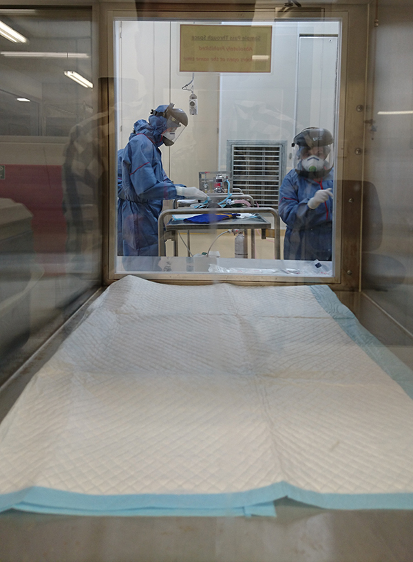
Being a Singapore-based research group (Duke-NUS) working in Australia, we needed facilities with enough space, resources and ideally sterile conditions and Bio Safety Cabinets. The University of Queensland (UQ) Veterinary School at Gatton, combined with its Queensland Animal Sciences Precinct (QASP) biosecurity facility, was ideal. It has a dedicated necropsy room designed for large animal quarantine purposes and attached Bio-Safety Level 2 (BSL2) tissue culture facilities (and BSL3 if required). As well as this, it gave us access to a supply chain including freezer and liquid nitrogen facilities, necessary for rapid freezing of cells and tissues. Of course, the ethical, government and legal paperwork took a while to sort out but thanks to great collaborators we got it all in place.
In the end, we had a platform for temporarily housing bats, quality sample collection, processing, cell suspension preparation and rapid storage of samples. We could then certify the samples were free of some specific pathogens (with help from the Australian Animal Health Laboratory), generate CITES permits and ship to an appropriate location. The team at UQ/QASP/CRC and all the staff from Duke-NUS/NUS/ Agency for Science, Technology and Research (A*STAR) that came for various bat trips were essential for making it all work. We know that not all researchers will have these resources available, but we hope our recommendations help influence their planning decisions.
Bat Surgery – Veterinarians Know Best
Having an expert veterinarian (Hume Field) and long-term bat handler (Gary Crameri) was key to the project’s success. These guys knew the intricacies of handling bats and were experienced enough to prevent any unnecessary wastage of precious animals. Of course, we threw them many challenges: asking them to identify specific lymph nodes, sections of the intestine; collecting stomach contents while not contaminating close by organs; and requiring a specific order of necropsy to prevent certain immune compartments (e.g. spleen/blood) from becoming activated due to time delay post-euthanasia. Even these experts were challenged with identification of the correct organs at first. Their experience was then passed on to our whole team and we felt the need to portray this knowledge to others.
Many research teams are now focusing on bats and it’s important for the field to develop consistent techniques in this less-common animal model. We also felt responsible for the use of such an ‘exotic animal’ and wanted to capitalise as much as we could to minimise the need for further captures. We hope that our videos (see the Supporting Information of our article) will help others in the field more clearly identify the intricacies of the necropsy (of course our GoPro is tough enough to resist the sterilization with Virkon!).
The Resources – Collaboration Welcome
There are already several bat cell-lines, but we’ve been able to generate many new cell lines for flying foxes and our research colony species,the cave nectar bat. In addition, we now have many primary cell lines (raised from multiple individuals of different ages/sexes etc), primary immune cell suspensions and bone marrow raised for differentiation or applicable for bat-mouse chimera studies..All of the organs collected were banked as cell suspensions and/or for tissue for DNA/RNA and protein with many having been processed already. Urine, faeces, sera etc are also available.

Some animals were part of larger experiments, so may be treated with immune stimulants. Others were collected as a great resource for population genetic studies and molecular evolution. Where possible, we recorded as much biological trait data as we could, with hopes of further meta-analysis in combination with molecular data. The animals were spread across estimated age groups and sexes and we may have tooth-aging data for some available soon.
We’ve already used these samples to highlight how bats have elevated expression of Heat Shock Proteins & therefore resistance to heat (though not drought), throughout their tissues. Along with this we identified elevated expression of ABCB1 (or MDR/P-Glycoprotein). This allows rapid removal of genotoxic compounds and may help explain the lack of cancerous tumors found in wild bats. Transcriptomic data from multiple individuals across many different organ types have been generated, too. This data is being used and overlaid with proteomic data or other validation for multiple projects. These cell lines and primary sources may be turned into in vitro models to prevent unnecessary captures in the future and we hope other researchers can discuss such opportunities with us – please get in touch.
To find out more about, read the full Methods in Ecology and Evolution Practical Tools article ‘Optimizing dissection, sample collection and cell isolation protocols for frugivorous bats’ (No Subscription Required)
