Post provided by Michelle Shero

To survive and reproduce, all animals must be able to extract sufficient energy from their environment. It takes energy to forage, but animals can recover those calories if they can successfully capture enough prey – and the animal’s weight tells us about its net energetic costs versus gains. Animals that remain in positive energy-balance can then afford to devote more energy towards growth and reproduction. In this blog post, Michelle Shero of Woods Hole Oceanographic Institution guides us through a new method outlined in her team’s recent Methods in Ecology and Evolution paper ‘Tracking wildlife energy dynamics with unoccupied aircraft systems and three-dimensional photogrammetry’. The team uses drone imagery to 3-D model and ‘weigh’ large groups of free-living animals.
What’s in a weight?
In most of her previous work, Shero would study a handful of animals for more comprehensive assessments of animal physiology and health including variation in lipid stores, intra-annual changes in stress and metabolic hormones, reproductive phenology, and (for marine mammals) dive capacities. Across the field of eco-physiology, researchers have found that almost all these critical aspects of the animal’s life history (and then some!) are correlated with body mass.
Weighing wild animals takes a lot of muscle power
On a typical project, these research teams were able to handle around 20-25 animals over 4-6 weeks of field work. That’s because it takes time to find and capture the animals, and physiological health assessments typically require that the animals be sedated. And physically weighing animals is hard! – Grey seals can weigh up to ~550 lbs (250 kg), and other species can weigh even more. Taking these traditional approaches on the ground only allows for a glimpse into the physiological status of a very small portion of the population. Furthermore, studies that involve animal handling or sedation often necessitate avoiding animals that are obviously malnourished or those that are visibly injured or have infections. This is for ethical reasons, to avoid adding any stressors to already-unhealthy animals, but it leaves a knowledge-gap in our understanding of underlying pathology in this important cohort of wild animals. We need to be able to measure the energy dynamics of animals more broadly across the population if we are to be able to truly assess the impacts of climate change and employ effective conservation measures.

‘Weighing’ more, with less
There has long been the interest in developing non-invasive ways to measure animal body size and mass. Previously, researchers have used hand-measured morphometrics to reconstruct the animal’s body shape using a series of elliptical truncated cones to calculate their body volume. For marine mammals, this can be paired with non-invasive blubber depth measurements taken from an ultrasound machine. This allows the morphometric models to be further deconstructed into blubber and ‘core’ body segments. Blubber masses calculated using these minimally-invasive methods have been previously compared with other ‘golden standards’ for measuring body lipid stores – namely using labeled water dilution techniques or proximate analysis. The high energetic-density of lipids makes this physiological metric a particularly valuable proxy of animal health.
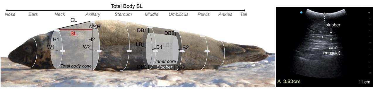
The desire to ‘weigh’ animals easily then led to the field of photogrammetry. On the ground, researchers could take side-profile or head-on photographs to measure the animal’s body length and width. Being able to model the animals in 3D provides a lot more dimensional information with which to measure fluxes in an animal’s caloric intake. Stereo-imagery for 3D modeling can be achieved with multiple researchers taking time-synchronous photographs around the animal’s body. Or a researcher can take a series of sequential photographs while walking 360-degrees around the animal to create ‘structure-from-motion’. On the ground, these methods required that some known-size objects be in the photograph’s field of view so that the final 3D model could be put to scale and the animals could be measured. Thus, this still required that people approach and disturb the animals, and animals had to be imaged one at a time.
Why not from the air?
There have been rapid technological advances using unoccupied aerial systems (UAS, or ‘drones’) in recent years. UAS are now commonly used to perform population counts, and researchers quickly realized the value in using UAS for 2D morphometrics using a single nadir photograph of the animals. This was ideal for garnering health indices for mobile and often difficult to access animals, such as cetaceans. However, the relationship between 2D measures and body volume varies by species, sex, age, and seasons. This often means that very specific calibration factors have to be developed for each study cohort. But with 3D models, animal body volume is a valuable measurement of body size in-and-of-itself, no matter what species or system you are studying. With so much success utilizing 3D photogrammetry on the ground — why couldn’t you do this from the air? The goal of Shero et al.’s study in Methods in Ecology and Evolution, was to use structure-from-motion 3D modeling techniques using aerial drones – and to do this on whole groups of animals at once. During flight, UAS collect GPS coordinates and altitude measurements. Because each image is georeferenced, the distance between photographs is known, and it is no longer necessary for researchers to walk up to the animals to place known-size objects in the photographs. Animals can be 3D modeled and ‘weighed’ from afar. This will allow us to ‘weigh’ more animals than would ever be possible from the ground, and makes strides towards weighing whole populations.
We sought to not only validate the new photogrammetric method, but we also wanted to show it ‘in action’ and actually demonstrate its applicability in answering larger ecological questions. We have carefully validated our new method by imaging groups of animals for 3D modeling, and then measuring and weighing some of those some individuals right afterwards for direct comparison. Aerial and ground measurements of seal body volume were equivalent. The UAS-derived 3D models could be used to estimate animal body mass with low error (2.1% body mass for adults; 9.8% for small pups).
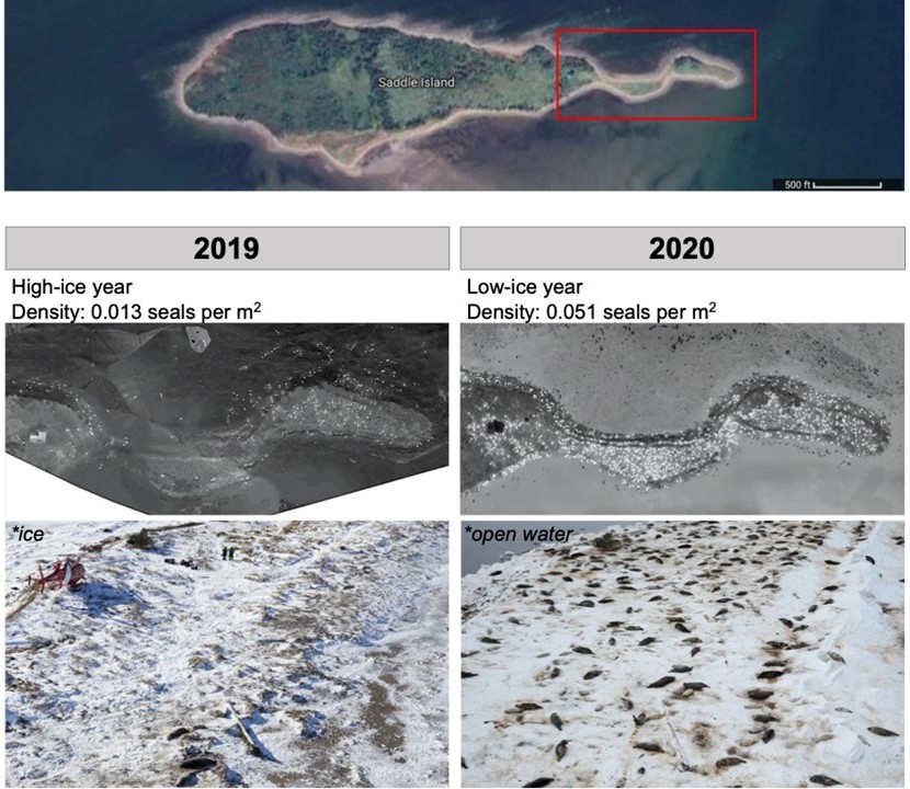
Our first field season was in January of 2019, and when we returned in 2020 we were stunned at how different the breeding colony looked. In 2019, there was a lot of local sea ice and, as we flew our helicopter over the area, we would see seals with their newborn pups on the ice substrate. At the breeding colony, there were very few seals spread out on-land. This was in stark contrast to 2020. We arrived at the breeding colony on our first day, only to find that were was no sea ice and the only place for the seals to give birth to their pups was on the island. There were orders of magnitude more seals than the previous year. The UAS 3D method was applied to almost 700 seals and revealed that female-to-pup energy transfer during lactation was significantly lower during the low-ice year in 2020 when animals were forced to give birth in very dense aggregations. This on-demand remote sensing provides a powerful tool with which to begin understanding how organismal level processes translate to population dynamics under varying environmental conditions. Moving forward, we hope that others will apply and adapt this non-invasive method to new wildlife systems for population monitoring.
To read the full study by Shero et al., “Tracking wildlife energy dynamics with unoccupied aircraft systems and three-dimensional photogrammetry”, visit the Methods in Ecology and Evolution journal website here.
To watch footage of UAS photogrammetry data collection from the seal colony, visit the YouTube video here.
